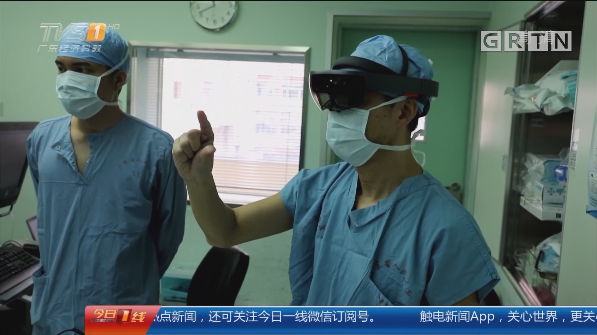
Abstract: Abstract: With the prevalence of deep neural networks, machine intelligence has recently demonstrated performance comparable with, and in some cases superior to, that of human experts in medical imaging and computer assisted intervention. Such accomplishments can be largely credited to the ever-increasing computing power, as well as a growing abundance of medical data. As larger clusters of faster computing nodes become available at lower cost and in smaller form factors, more data can be used to train deeper neural networks with more layers and neurons, which usually translate to higher performance and at the same time higher computational complexity. For example, the widely used 3D U-Net for medical image segmentation has more than 16 million parameters and needs about 4.7×10¹³ floating point operations to process a 512×512×200 3D image. The large sizes and high computation complexity of neural networks have brought about various issues that need to be addressed by the joint efforts between hardware designers and medical practitioners towards hardware aware learning. In this talk, I will present novel solutions for the data acquisition and data processing stages in medical image computing respectively, using hardware-oriented schemes for lower latency, memory footprint and higher performance in embedded platforms. I will discuss how our hardware-aware machine learning approaches led to the real-time MRI segmentation for prosthetic valve implantation assistance, and enabled the world’s first AI assisted telementoring of cardiac surgery on April 3, 2019.
Abstract: The increasing prevalence of chronic diseases, an aging population, and a shortage of healthcare professionals have prompted the widespread adoption of mobile and implantable devices to effectively manage various health conditions. In recent years, there is growing interest to leverage the rapid advances in artificial intelligence (AI) to enhance the performance of these devices, resulting in better patient outcomes, reduced healthcare costs, and improved patient autonomy. Due to privacy, security, and safety considerations, inferences must often be done on the edge, with limited hardware resources. This is compounded by inter-patient and intra-patient variability, heavy dependence on medical domain knowledge, and lack of diversified training data. In this talk, we will demonstrate how techniques such as hardware and neural architecture co-design, personalized meta-learning, and fairness-aware pruning can transform the landscape of mobile and implantable devices. Additionally, we will showcase the world's first smart Implantable Cardioverter Defibrillator (ICD) design enabled by our research..
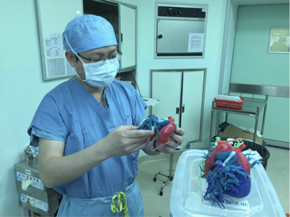
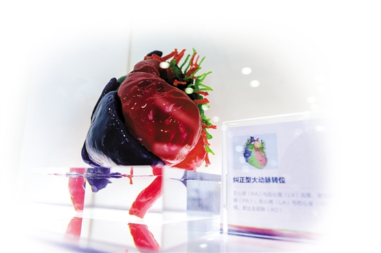
2021年11月25日庄建医生是广东省人民医院心血管医学3D打印实验室主任。 在进行先天性心脏病手术前,他和团队可以把病人心脏模型打印出来,缩减手术判断时间、 提升治疗效果。目前,他已经将3D打印技术应用于数百例病人的术前规划。顾名思义, 3D打印技术不是用油墨在纸张上打印内容,而是在三维空间里逐层打印出立体的东西。 这一新兴技术正加速在我国医疗领域应用,落地场景日渐广泛。“新华视点”记者了解到, 包括北京大学第三医院、北京积水潭医院、南方医科大学第三附属医院等在内的多家医院, 已将其运用于术前规划、手术导板、人体植入等。

2018年6月25日,来自美国圣母大学和华中科技大学的研究者们提出 了一种利用网络量化提升用于医疗影像分割的深度学习模型精度的优化方法。 该方法创新性地将旨在压缩网络模型的量化方法应用到医疗影像分割深度学习 模型中,相比于已有的方法,针对当前流行的 Gland 数据集可提升当前 state-of-the-art 的图片分割准确度达 1%-7.5%, 同时获得压缩了的深度学习模型。该研究已被 CVPR 2018 接收。
报道媒体: 搜狐
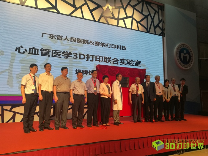
2017年10月13日,这是由省人民医院、省心血管病研究所和珠海赛纳打印科技股份有限公司成立的国内 首个心血管医学3D打印医研企联合实验室,旨在推动心血管医学3D打印技术的临床应用、 科学研究和国产化3D打印设备与材料的研发。记者了解到, 实验室目前将承担利用3D打印模型协助医生做术前诊断、术中“导航”和术后评估等; 预计两年内,可以实现个体化定制打印体内可植入的生物或非生物移植物;未来有望造出可供移植的“人造心脏”。
报道媒体: 新华网